Citation
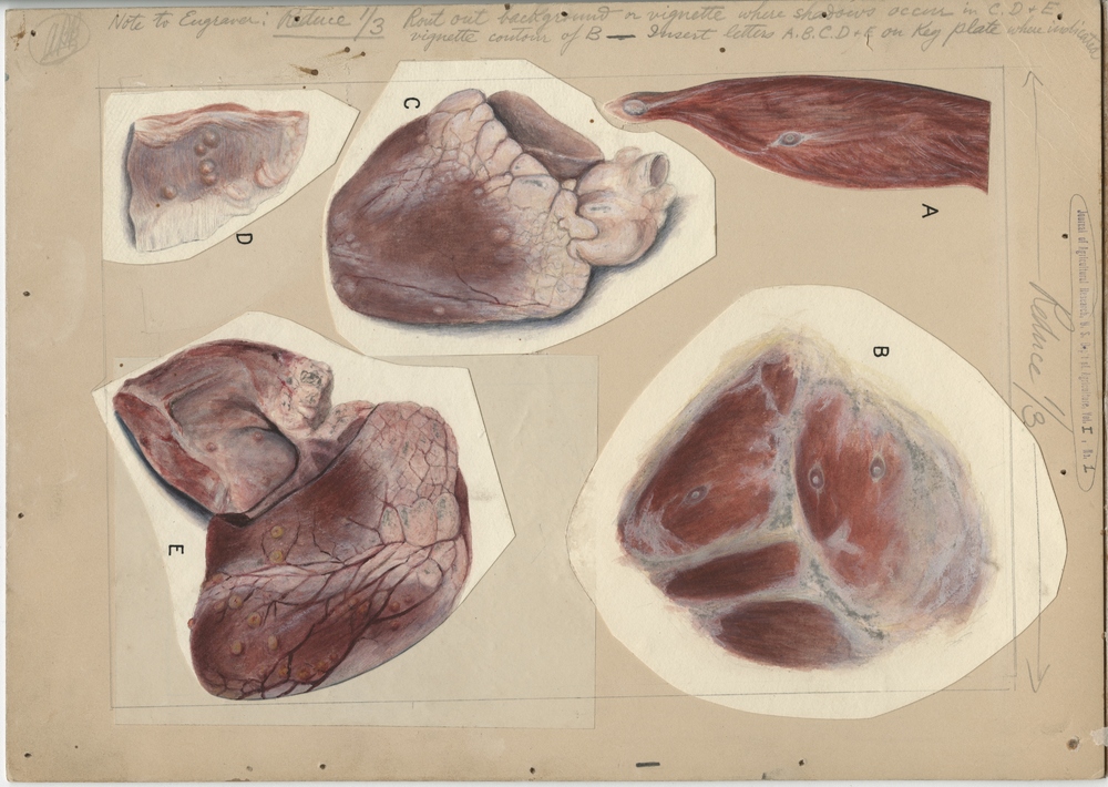
William S.D. Haines. 1913. “Figures A and B - Portions of muscle of sheep showing Cysticercus ovis (undegenerated) in situ. Figure A. - Section of hind leg showing two "deep" cysticerci. Figure B. - Hind leg showing three "superficial" cysticerci. Figures C and D - Heart and portion of diaphragm of sheep showing Sarcocystis nodules likely to be mistaken for degenerate cysticerci. Figure E. - Sheep heart showing numerous small degenerate cysticerci (Cysticercus ovis). Illustration for Journal of Agricultural Research, Volume 1, Number 1, Plate III..” Special Collections, USDA National Agricultural Library. Accessed April 25, 2024, https://www.nal.usda.gov/exhibits/speccoll/items/show/8087.
 An official website of the United States government.

